Radiodiagnosis is the branch of nuclear medicine that deals with diagnosing and treating diseases using information and images of the patient using ionizing or non-ionizing radiation and different types of energy.
Over the years, it has become an essential service for patient care. Through the images captured from the inside of different body parts, doctors can verify the progress of a disease in a systematic study. As a result, they can detect abnormalities, diagnose, and determine treatments for injuries or illnesses.
Radiodiagnostic tests consist of obtaining images for the diagnosis of the interior of the organism using X-ray equipment, which crosses the field to be studied. The most common diagnostic radiology studies include mammograms, plain x-rays, ultrasounds or sonograms, and computed tomography. The Imaging Centers at Salus clinics offer X-ray studies, CT scans, general sonography, Dexa, MRI, and mammograms.
This branch of medicine is an example of the use of nuclear energy in the civil sphere to diagnose and treat diseases. Numerous advances have been made in this field, highlighting several techniques that do not use ionizing radiation, such as MRI and ultrasound.
What is radiology?
Medical radiology deals with the production and interpretation of images obtained through the use of ionizing radiation. Radiologists are doctors of medicine who have received specialized training in this medical science.
This concept is sometimes also called diagnostic radiology or radiodiagnosis.
Radiology can be divided into two different areas diagnostic radiology and interventional radiology:
-
Diagnostic radiology: radiologic technologists use this technique to see structures inside the body and to be able to diagnose the causes of specific symptoms or detect diseases.
-
Interventional Radiology: Serves to help guide medical procedures. These images allow interventional radiologists to insert probes, instruments, or small tools into the body. They are mainly used to detect and treat cancer in almost any body part.
Radiology in children
Pediatric radiology is the branch of radiology that deals with diagnostic imaging in the pediatric field. This specialty arises from the need to carry out radiological research in children with different criteria from adults due to obvious protectionist problems.
Radiodiagnostic techniques
Radiology uses X-rays for the production of medical images, but not solely. The main techniques used by radiodiagnosis doctors to obtain radiological images are the following:
-
Conventional radiography
-
Computerized axial tomography (CT).
-
Positron emission tomography (PET)
-
Ultrasound and sonography
-
Magnetic resonance imaging (MRI)
Ultrasound and MRI do not require ionizing radiation.
Thanks to X radiology, a radiology technician can perform studies inside the human body, including, for example, the thorax, abdomen, skeleton, digestive tract, heart, thorax, abdomen, nervous system, and urinary system, among others.
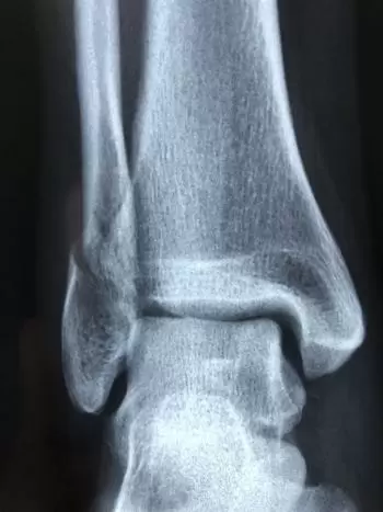
Conventional radiography
It is the oldest technique but is still used for specific diagnoses. In it, a radioactive source emits energy that the tissues absorb. The particles that the tissues do not stop are captured on a plate of photographic paper.
The result of the photograph is that the hard tissues appear white in the photo, and the darker parts represent the softer tissues.
This test is indicated in most diagnostic imaging studies since it emits very little radiation and is helpful for diagnosing some pathologies.
Pros of diagnosing using x-rays:
-
It helps in a non-invasive and painless way to diagnose diseases and control treatments and therapies.
-
Support the planning of medical and surgical treatment, before, after, and even in some cases, during the intervention.
-
Guide medical personnel as they insert catheters, stents, or other devices into the body, treat tumors or remove blood clots or other blockages.
Cons of the use of x-rays
As with many aspects of medicine, there are risks associated with using X-ray imaging, which uses ionizing radiation to create images of the body. Ionizing radiation is a form of radiation that has enough energy that, at certain powers, it causes DNA damage if usually tightly controlled limits are exceeded.
These risks include:
-
There is a slight increase in the chance that a person exposed to X-rays will develop cancer. It depends on the type of procedure, the amount of exposure, and the number of times you undergo this procedure per year. This is why radiology personnel must be protected because they are the most exposed. In general, the patient does not have to worry since medical protocols establish safety margins that guarantee not to harm her health.
-
Another risk of X-ray imaging is possible reactions associated with an intravenously injected contrast agent, sometimes used to enhance visualization.
Computed tomography (CT)
Computerized axial tomography (CT) consists of obtaining a 3D projection of an organ on a computer. This 3D image is obtained by computer from the images of the superimposed cuts of the organ to be studied. The different images are obtained through a finely collimated X-ray beam that rotates around it.
The level of radiation that the patient receives in a CT scan is higher than that of radiography.
Positron emission tomography (PET)
Doctors can see how your organs and tissues work with positron emission tomography. For them, a radioactive substance marker is used to show the structure and blood flow through the organs.
Sonography
 Ultrasounds are performed by sending ultrasound to the part of the body under study, having placed a sound-conducting gel. The transducer collects the ultrasound that will shape the body's internal structure that is being studied through a screen in real time.
It is the most minimally invasive imaging technique for the human body since it does not emit any type of radiation. For this reason, it is even performed on pregnant women to study the state of the fetus.
Magnetic resonance imaging MRI
Magnetic resonance imaging consists of using a magnetic field, a radio wave emitter and receiver, and a computer to obtain images of tissues. It is performed by introducing a tube-shaped camera to study the area. Then, with the action of the magnetic field, the tissues respond by modifying the emitted radio signal.
How is an MRI performed?
During the MRI, the patient will lie inside the machine (although there are open and closed ones, it is usually similar to a long, narrow tube, approximately one meter in diameter and opened at the ends).
Inside the tube, the person will be surrounded by a magnetic field that reacts with the magnetic elements inside the body and transmits a weak radio signal. This signal is collected and processed in a computer, offering an image that can be displayed on the screen. The most annoying part of this test is a loud and monotonous noise generated by the machine itself, which is why some professionals recommend listening to music to minimize the impact.
Meanwhile, the imaging technician will be in an adjacent room from which he will control the patient at all times and will be able to talk to him through an intercom.
Finally, during the entire test, the patient must remain immobile so that the results are as precise as possible.
MRI risks
Although, as we have already detailed, magnetic resonance is a very safe test since it uses a powerful magnetic field to obtain the images. People with electronic devices or any metallic component in their body should inform the specialist before starting the test to ensure their safety.