Nuclear medicine is a branch of medicine that uses radionuclides, also known as radioisotopes, for the diagnosis, treatment and monitoring of various medical conditions.
These radioactive elements have proven to be valuable tools in the field of health, allowing medical professionals to obtain crucial information about the functioning of the human body.
In this article, we will explain in detail radionuclides in nuclear medicine, their applications, risks and benefits.
What are radionuclides?
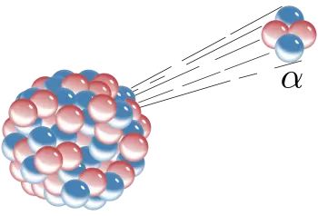
Radionuclides, or radioisotopes, are atoms that have an unusual number of neutrons in their nucleus, making them unstable and radioactive. This instability manifests itself through the emission of subatomic particles and/or energy in the form of radiation. These radioactive properties are what make radionuclides valuable in nuclear medicine.
In nuclear medicine, radionuclides with short or moderate half-lives are used. The half-life is the time it takes for half of a quantity of radionuclides to decay.
This allows radionuclides to be active enough to provide useful medical data, but not so active as to pose a significant health risk.
Applications in medicine
Radionuclides are used in nuclear medicine in a variety of applications, the most common being:
Diagnostic imaging
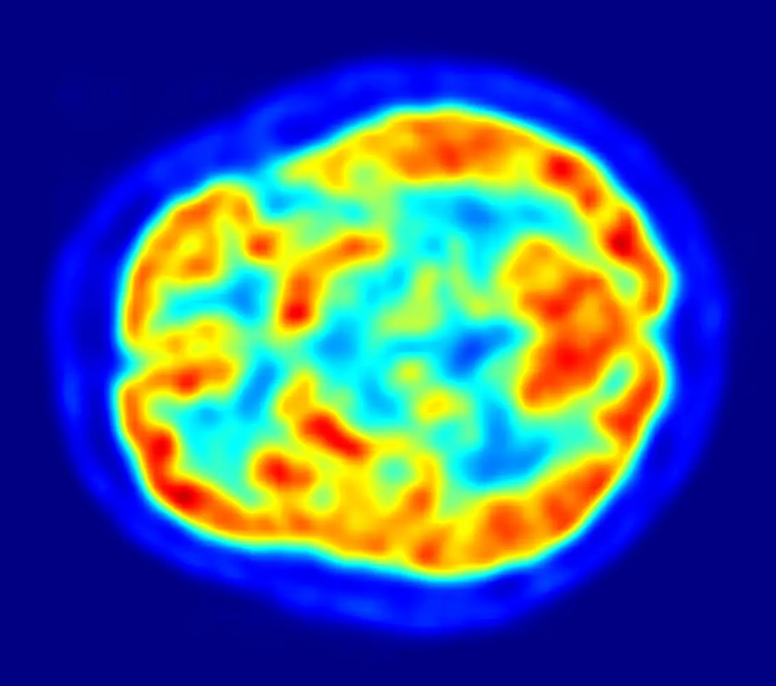
Scintigraphy and positron emission tomography (PET) are two techniques that use radionuclides to obtain detailed images of the inside of the body.
In scintigraphy, a radioactive radionuclide is given to the patient and accumulates in a specific part of the body. The emitted radiation is detected and converted into images that allow doctors to diagnose diseases such as cancer, heart disease and bone disorders.
Cancer treatment
Radionuclide radiation therapy is used to treat cancer.
In this case, controlled doses of radionuclides are delivered directly to the cancer cells, helping to destroy them. Iodine-131, for example, is used to treat thyroid cancer, while lutetium-177 is used for neuroendocrine cancer.
Organ function studies
Radionuclides are also used to evaluate the function of specific organs. An example is the study of kidney function through the use of technetium-99m, which is combined with substances that the kidney filters and excretes. The amount of radionuclide in the urine provides information about kidney function.
Bone pain therapy
In cases of bone metastases and associated pain, radionuclides such as strontium-89 and samarium-153 are used to relieve pain and reduce inflammation in the affected bones.
Safety Risks and Considerations
Although radionuclides are valuable tools in nuclear medicine, their use is not without risks and safety considerations. Some of these risks include:
Radiation
Radiation emitted by radionuclides is a major concern. Patients and the medical staff who work with them are exposed to radiation. However, doses are kept as low as possible and strict safety protocols are followed to minimize exposure.
Secure deletion
The safe disposal of radioactive materials is essential to prevent environmental contamination. Special containers are used and strict regulations are followed for the disposal of radioactive waste.
Normative compliance
Government regulation and oversight are essential to ensure the safe use of radionuclides in nuclear medicine. Medical centers and laboratories that use radionuclides must comply with local and international laws and regulations.
Precautions in pregnancy and breastfeeding
It is essential to take extra precautions with pregnant or nursing patients, as radiation may be harmful to the fetus or infant.
Examples of radionuclides for nuclear medicine
 In nuclear medicine, a variety of radionuclides are used for diagnostic and therapeutic applications.
Below are some of the most commonly used radionuclides in nuclear medicine:
- Technetium-99m (99mTc): Technetium-99m is used in scintigraphy to obtain images of various organs and tissues, such as the heart, brain, bones, and kidney system. Its short half-life allows high-quality images to be obtained without prolonged exposure to radiation.
- Iodine-131 (131I): Iodine-131 is used in the treatment of thyroid cancer, as iodine is absorbed by thyroid tissue. The radiation emitted by iodine-131 destroys abnormal thyroid cells.
- Fluorine-18 (18F): Fluorine-18 is used in positron emission tomography (PET), which allows the detection of tumors and other metabolic conditions in the body. It is commonly used in combination with radiolabeled glucose (FDG) to visualize metabolic activity.
- Cobalt-60 (60Co): Cobalt-60 is used in radiation therapy to treat tumors. Its high energy allows deep tissue penetration, making it effective in the treatment of cancer.
- Iodine-123 (123I): Iodine-123 is used in thyroid scintigraphy and in studies of thyroid gland function. It has a shorter half-life compared to iodine-131 and is used for diagnostic imaging.
- Gallium-67 (67Ga): Gallium-67 is used in scintigraphy for the detection of inflammation and abscesses in the body. It is also used in some cases to detect tumors.
- Lutetium-177 (177Lu): Lutetium-177 is used in radionuclide therapy to treat certain types of cancer, such as neuroendocrine cancer and metastatic prostate cancer.
- Thallium-201 (201Tl): Thallium-201 is used in cardiac scintigraphy to evaluate heart function and blood flow. It is particularly useful in the detection of cardiac ischemia.
- Strontium-89 (89Sr): Strontium-89 is used in the treatment of bone pain in cases of bone metastases, as it accumulates in the affected bone tissue.
- Samarium-153 (153Sm): Samarium-153 is also used in the treatment of bone pain in patients with bone metastases. Emitting beta radiation, it acts directly on the affected areas.
Medical benefits of radionuclides
Despite the risks associated with the use of radionuclides, the benefits in nuclear medicine are undeniable. These are some of the most significant advantages:
- Diagnostic accuracy : Nuclear medicine allows for accurate, non-invasive assessment of organ and tissue function and structure, helping doctors make more informed treatment decisions.
- Effective Cancer Treatment : Radionuclide therapy has been shown to be effective in treating certain types of cancer, offering patients a valuable therapeutic option.
- Personalization of treatment : Nuclear medicine allows the personalization of treatments and adaptation to the specific needs of each patient.
- Advanced medical research : Radionuclides are also essential in medical research, contributing to advances in knowledge and the development of new therapies and diagnostics.